Hep2 Cell Patterns
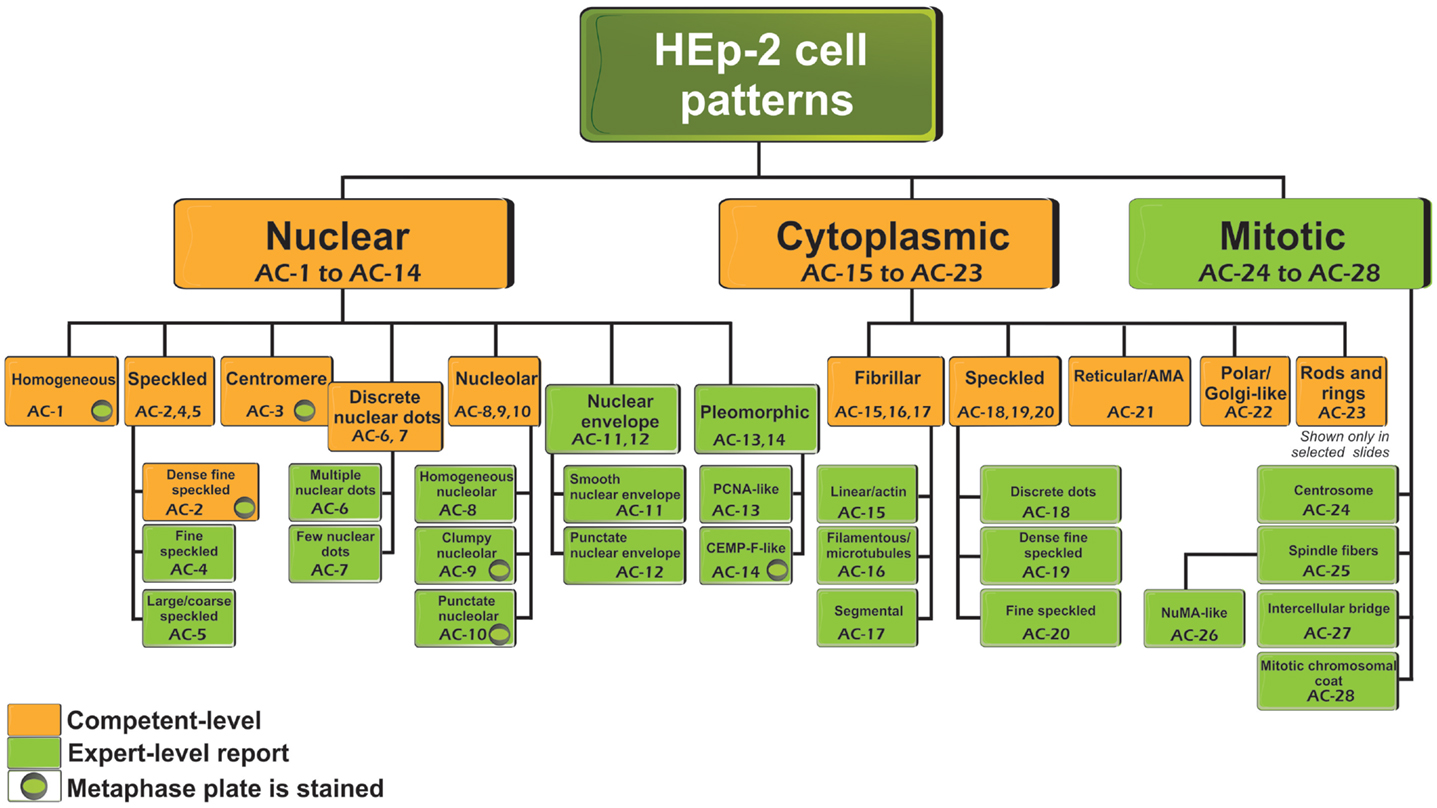
Hep2 Cell Patterns - Homogenous, speckled, centromere, nucleolar, and nuclear dots. This clinical relevance is primarily defined within the context of the suspected disease and includes recommendations for. We conclude hereby that synucleinopathies are not associated with detectable presence of ana in plasma. It still leaves open the question of. Interphase cells show homogeneous nuclear staining while mitotic cells show staining of the condensed chromosome regions. Nuclear homogeneous, nuclear coarse speckled, and nuclear centromeric patterns appeared exclusively in patients with ards. Serum complement 3 (c3), c4, and immunoglobulin g were compared among subgroups with different ana titers. This is a summary of the international consensus on antinuclear antibody pattern (icap) meeting and subsequent discussion, debate, and dialog. The dichotomous outcome, negative or positive, is integrated in diagnostic and classification criteria for. Many patients with sle have more than one type of pattern. Serum complement 3 (c3), c4, and immunoglobulin g were compared among subgroups with different ana titers. This clinical relevance is primarily defined within the context of the suspected disease and includes recommendations for. Web assess antinuclear antibody titers and patterns were retrospectively identified and compared by iifa using human epithelial cells (hep‐2) and primate liver tissue substrate according to international consensus in sard. Homogenous, speckled, centromere, nucleolar, and nuclear dots. These patterns are the result of autoantibody binding. International consensus on ana patterns. Interphase cells show homogeneous nuclear staining while mitotic cells show staining of the condensed chromosome regions. Web it allows detection of antibody binding to specific intracellular targets, resulting in diverse staining patterns that are usually categorized based on the cellular components recognized and the degree of binding, as reflected by the fluorescence intensity or titer [ 2, 3 ]. This is a summary of the international consensus on antinuclear antibody pattern (icap) meeting and subsequent discussion, debate, and dialog. Many patients with sle have more than one type of pattern. This clinical relevance is primarily defined within the context of the suspected disease and includes recommendations for. Nuclear homogeneous, nuclear coarse speckled, and nuclear centromeric patterns appeared exclusively in patients with ards. These patterns are the result of autoantibody binding. The dichotomous outcome, negative or positive, is integrated in diagnostic and classification criteria for. Interphase cells show homogeneous nuclear staining. Experienced cl defined as reporting all 3 main nomenclature categories. Web it allows detection of antibody binding to specific intracellular targets, resulting in diverse staining patterns that are usually categorized based on the cellular components recognized and the degree of binding, as reflected by the fluorescence intensity or titer [ 2, 3 ]. Homogenous, speckled, centromere, nucleolar, and nuclear dots.. Web the ana pattern profile was distinct in the 2 groups. Experienced cl defined as reporting all 3 main nomenclature categories. These patterns are the result of autoantibody binding. The consensus paper has been published in annals of the rheumatic diseases.1. Interphase cells show homogeneous nuclear staining while mitotic cells show staining of the condensed chromosome regions. These patterns are the result of autoantibody binding. This is a summary of the international consensus on antinuclear antibody pattern (icap) meeting and subsequent discussion, debate, and dialog. It still leaves open the question of. Many patients with sle have more than one type of pattern. Nuclear homogeneous, nuclear coarse speckled, and nuclear centromeric patterns appeared exclusively in patients with. This clinical relevance is primarily defined within the context of the suspected disease and includes recommendations for. The consensus paper has been published in annals of the rheumatic diseases.1. It still leaves open the question of. Many patients with sle have more than one type of pattern. Interphase cells show homogeneous nuclear staining while mitotic cells show staining of the. These patterns are the result of autoantibody binding. Web the ana pattern profile was distinct in the 2 groups. The consensus paper has been published in annals of the rheumatic diseases.1. Interphase cells show homogeneous nuclear staining while mitotic cells show staining of the condensed chromosome regions. Web assess antinuclear antibody titers and patterns were retrospectively identified and compared by. The dichotomous outcome, negative or positive, is integrated in diagnostic and classification criteria for. This clinical relevance is primarily defined within the context of the suspected disease and includes recommendations for. Nuclear homogeneous, nuclear coarse speckled, and nuclear centromeric patterns appeared exclusively in patients with ards. We conclude hereby that synucleinopathies are not associated with detectable presence of ana in. The consensus paper has been published in annals of the rheumatic diseases.1. Interphase cells show homogeneous nuclear staining while mitotic cells show staining of the condensed chromosome regions. Many patients with sle have more than one type of pattern. It still leaves open the question of. These patterns are the result of autoantibody binding. Web it allows detection of antibody binding to specific intracellular targets, resulting in diverse staining patterns that are usually categorized based on the cellular components recognized and the degree of binding, as reflected by the fluorescence intensity or titer [ 2, 3 ]. This clinical relevance is primarily defined within the context of the suspected disease and includes recommendations for.. The nuclear dense fine speckled pattern occurred only in healthy individuals. Interphase cells show homogeneous nuclear staining while mitotic cells show staining of the condensed chromosome regions. Web assess antinuclear antibody titers and patterns were retrospectively identified and compared by iifa using human epithelial cells (hep‐2) and primate liver tissue substrate according to international consensus in sard. Web the ana. The nuclear dense fine speckled pattern occurred only in healthy individuals. These patterns are the result of autoantibody binding. It still leaves open the question of. Interphase cells show homogeneous nuclear staining while mitotic cells show staining of the condensed chromosome regions. The consensus paper has been published in annals of the rheumatic diseases.1. Serum complement 3 (c3), c4, and immunoglobulin g were compared among subgroups with different ana titers. This clinical relevance is primarily defined within the context of the suspected disease and includes recommendations for. Homogenous, speckled, centromere, nucleolar, and nuclear dots. We conclude hereby that synucleinopathies are not associated with detectable presence of ana in plasma. Many patients with sle have more than one type of pattern. Web the ana pattern profile was distinct in the 2 groups. The dichotomous outcome, negative or positive, is integrated in diagnostic and classification criteria for. This is a summary of the international consensus on antinuclear antibody pattern (icap) meeting and subsequent discussion, debate, and dialog. Web assess antinuclear antibody titers and patterns were retrospectively identified and compared by iifa using human epithelial cells (hep‐2) and primate liver tissue substrate according to international consensus in sard.
HEp2 staining patterns 1) Homogeneous 2) Nucleolar 3) Coarse Speckled

The surface of six Hep2 cell patterns. Download Scientific Diagram

Figure 1 from The Classification of HEp2 Cell Patterns Using Fractal

Representative images of selected major HEp2 cell patterns. (A

2. IFA Pattern recognition & HEp2 cell components YouTube

Representative images of selected major HEp2 cell patterns. (A
Frontiers Report of the First International Consensus on Standardized

Display of HEp2 cell pattern classification agreement and disagreement

Figure 1 from The Clinical Significance of the Dense Fine Speckled

Frontiers Report of the First International Consensus on Standardized
International Consensus On Ana Patterns.
Web It Allows Detection Of Antibody Binding To Specific Intracellular Targets, Resulting In Diverse Staining Patterns That Are Usually Categorized Based On The Cellular Components Recognized And The Degree Of Binding, As Reflected By The Fluorescence Intensity Or Titer [ 2, 3 ].
Experienced Cl Defined As Reporting All 3 Main Nomenclature Categories.
Nuclear Homogeneous, Nuclear Coarse Speckled, And Nuclear Centromeric Patterns Appeared Exclusively In Patients With Ards.
Related Post: